%20--%3e%3csvg%20version='1.1'%20id='Ebene_1'%20xmlns='http://www.w3.org/2000/svg'%20xmlns:xlink='http://www.w3.org/1999/xlink'%20x='0px'%20y='0px'%20viewBox='0%200%20897.61%20762.46'%20style='enable-background:new%200%200%20897.61%20762.46;'%20xml:space='preserve'%3e%3cstyle%20type='text/css'%3e%20.st0{fill:%2305171D;}%20%3c/style%3e%3cg%3e%3cpath%20class='st0'%20d='M211.66,376.86h-2.38c-7.13,27.08-34.67,47.52-66.99,47.52c-63.19,0-95.97-46.1-95.97-130.67%20c0-84.58,32.78-130.19,95.97-130.19c32.32,0,59.86,19.96,66.99,47.51h2.38V67.07h70.32v351.61h-70.32V376.86z%20M211.66,330.78%20v-73.64c0-22.81-19.48-36.59-46.09-36.59s-46.09,20.43-46.09,51.8v43.24c0,31.36,19.48,51.79,46.09,51.79%20S211.66,353.58,211.66,330.78'/%3e%3cpath%20class='st0'%20d='M352.28,223.87h-35.16v-54.64h35.16v-29.46c0-48,26.13-72.7,74.6-72.7h45.61v54.65h-49.9v47.51h49.9v54.64%20h-49.9v194.81h-70.32V223.87z'/%3e%3cpolygon%20class='st0'%20points='503.46,67.07%20573.78,67.07%20573.78,274.71%20576.63,274.71%20607.53,228.62%20657.41,169.23%20735.33,169.23%20652.19,264.73%20744.83,418.67%20661.21,418.67%20605.14,313.2%20573.78,348.36%20573.78,418.67%20503.46,418.67%20'/%3e%3crect%20x='773.37'%20y='169.22'%20class='st0'%20width='70.32'%20height='249.45'/%3e%3cpath%20class='st0'%20d='M808.53,52.95c20.96,0,38.01,17.05,38.01,38.01c0,20.96-17.05,38.01-38.01,38.01%20c-20.95,0-38.01-17.05-38.01-38.01C770.52,70,787.58,52.95,808.53,52.95%20M808.53,48.2c-23.61,0-42.76,19.15-42.76,42.76%20c0,23.61,19.15,42.76,42.76,42.76c23.61,0,42.76-19.15,42.76-42.76C851.29,67.35,832.14,48.2,808.53,48.2'/%3e%3cpath%20class='st0'%20d='M838.94,703.81h-60.82V463.87h60.82V703.81z%20M843.69,459.11h-70.32v249.45h70.32V459.11z'/%3e%3cg%3e%3cpath%20class='st0'%20d='M712.05,653.92V544.64c0-58.92-36.11-91.22-106.44-91.22c-52.26,0-80.77,18.05-100.72,47.51l41.81,37.06%20c10.93-15.2,25.66-29.46,53.69-29.46c29.46,0,41.33,15.2,41.33,39.43v15.68h-39.42c-66.06,0-106.91,25.19-106.91,77.93%20c0,43.24,27.56,72.69,77.44,72.69c35.05,0,63.89-13.96,74.36-39.92h2.38c6.68,21,22.97,34.22,44.93,34.22h40.83v-54.64H712.05z%20M730.58,703.81H694.5c-20.72,0-36.15-13.03-41.53-34.22h-1.73h-1.27h-0.82h-3.21h-1.77c-8.71,25.38-36.29,39.92-71.34,39.92%20c-45.52,0-72.69-25.39-72.69-67.94c0-60.48,55.55-73.18,102.16-73.18h39.42h4.75v-4.75v-15.68c0-28.91-15.94-44.19-46.09-44.19%20c-28.04,0-43.84,13.77-54.39,27.23l-34.78-30.83c21.11-29.03,50.43-42.02,94.4-42.02c66.53,0,101.69,29.9,101.69,86.47v109.29%20v4.75h4.75h18.53V703.81z'/%3e%3cpath%20class='st0'%20d='M646.47,603.56v-4.75h-4.75h-34.21c-40.1,0-46.09,20.54-46.09,32.78v7.13c0,11.71,4.87,31.36,37.53,31.36%20c29.3,0,47.51-14.21,47.51-37.06V603.56z%20M641.72,633.02c0,22.81-19.48,32.31-42.76,32.31c-20.9,0-32.78-8.55-32.78-26.61v-7.13%20c0-18.05,14.25-28.03,41.34-28.03h34.21V633.02z'/%3e%3c/g%3e%3c/g%3e%3c/svg%3e)
Expertise · Medical Image Analysis
Medical Image Analysis
AI-driven segmentation, classification and multimodal image analysis for diagnostics, therapy planning and clinical research.
Selected DFKI research projects
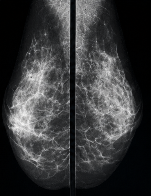
Mammography screening
AI-assisted analysis of screening mammograms for early breast cancer detection.
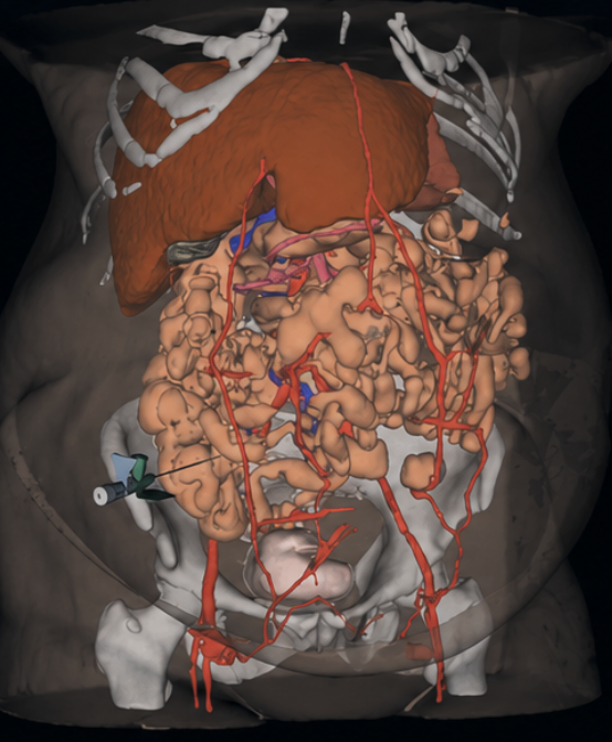
3D organ & vessel segmentation
Multi-organ and vascular segmentation from CT for surgical and therapy planning.
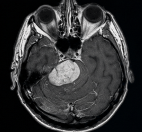
Neuro-oncological MRI
Lesion detection and characterisation on brain MRI for neuro-oncology.
Research Focus
Scientific excellence applied to real-world healthcare.
DFKI research in medical image analysis combines deep learning, computer vision and clinical domain knowledge to interpret imaging data across modalities — supporting radiology, pathology, ophthalmology and surgical planning through validated methods.
Methods and capabilities
- Semantic and instance segmentation
- Lesion, structure and tissue classification
- Multimodal image fusion (CT, MRI, ultrasound, histology)
- Self-supervised and weakly-supervised learning
- Uncertainty estimation and quality control
- Evaluation and benchmarking on clinical datasets
Application areas
- Radiology decision support
- Digital pathology
- Ophthalmic imaging
- Surgical and therapy planning
- Clinical research and cohort analysis
Engagements typically start with a feasibility study or joint research project — evaluating data, methods and impact before wider deployment.
Related Expertise & Services
Where this research connects.
Complex healthcare challenges rarely map to a single method. Our expertise areas combine to support broader clinical, research and product initiatives.
Medical AI R&D Partner
Research-to-prototype collaboration across imaging, sensing and movement analysis.
Learn moreMedical Image Registration
Longitudinal alignment for disease progression and therapy monitoring.
Learn moreExplainable Medical AI
Transparent, interpretable AI methods for clinical trust and validation.
Learn moreMedical AI Solutions
Applied medical AI across imaging, diagnostics and clinical workflows.
Learn moreImage analysis is frequently the core of new imaging products — learn how research moves from idea to AI-enabled medical device prototype.
Explore this research expertise with DFKI.
From feasibility studies to long-term research collaboration — start with a structured conversation about your challenge.
Stay connected with Healthcare AI research and innovation — Join the Community